Home > CSC-OpenAccess Library > Manuscript Information
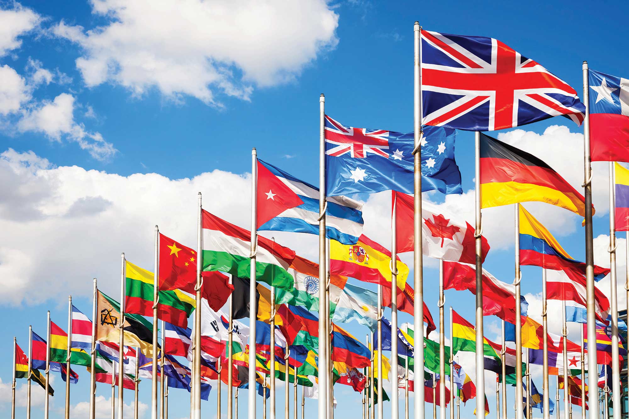
EXPLORE PUBLICATIONS BY COUNTRIES |
 |
| EUROPE | |
| MIDDLE EAST | |
| ASIA | |
| AFRICA | |
| ............................. | |
| United States of America | |
| United Kingdom | |
| Canada | |
| Australia | |
| Italy | |
| France | |
| Brazil | |
| Germany | |
| Malaysia | |
| Turkey | |
| China | |
| Taiwan | |
| Japan | |
| Saudi Arabia | |
| Jordan | |
| Egypt | |
| United Arab Emirates | |
| India | |
| Nigeria | |
Knee Joint Articular Cartilage Segmentation using Radial Search Method, Visualization and Quantification
M S Mallikarjuna Swamy, Mallikarjun S. Holi
Pages - 1 - 13 | Revised - 15-01-2013 | Published - 28-02-2013
MORE INFORMATION
KEYWORDS
Cartilage, Image Segmentation, Knee Joint, MRI, Osteoarthritis
ABSTRACT
Knee is a complex and highly stressed joint of the human body. Articular Cartilage is a smooth
hyaline spongy material between the tibia and femur bones of knee joint. Cartilage morphology
change is an important biomarker for the progression of osteoarthritis (OA). Magnetic Resonance
Imaging (MRI) is the modality widely used to image the knee joint because of its hazard free and
soft tissue contrast. Cartilage thickness measurement and visualization is useful for early
detection and progression of the disease in case of OA affected patients. In the present work,
knee joint MR images of normal and OA affected are processed for segmentation and
visualization of cartilage using semiautomatic method. The radial search method is used with
minor modifications in search area to reduce computation time. Cartilage thickness and volume is
measured in lateral, medial and patellar regions of femur. The overall accuracy of measurements
is determined by comparing the measurements with another semiautomatic method based on
edge detection and interpolation. It is observed a good correlation between quantification of
cartilage in two methods. The method takes less time for segmentation because of reduced
manual steps. The reduced cartilage thickness and volume is observed in OA affected knee of
different level of progression.
| 1 | Aprovitola, A., & Gallo, L. (2016). Knee bone segmentation from MRI: A classification and literature review. Biocybernetics and Biomedical Engineering. |
| 2 | Hani, A. F. M., Kumar, D., Malik, A. S., Walter, N., Razak, R., & Kiflie, A. (2015). Multinuclear MR and Multilevel Data Processing: An Insight into Morphologic Assessment of In Vivo Knee Articular Cartilage. Academic radiology, 22(1), 93-104. |
| 3 | Ahmad, J., Malik, A. S., Abdullah, M. F., Kamel, N., & Xia, L. (2015). A novel method for vegetation encroachment monitoring of transmission lines using a single 2D camera. Pattern Analysis and Applications, 18(2), 419-440. |
| 4 | Hani, A. F. M., Kumar, D., Malik, A. S., Ahmad, R. M. K. R., Razak, R., & Kiflie, A. (2015). Non-invasive and in vivo assessment of osteoarthritic articular cartilage: a review on MRI investigations. Rheumatology international, 35(1), 1-16. |
| 5 | Bakir, H., & Zrida, J. (2014, September). Automatic knee cartilage segmentation and visualization. In Advances in Computing, Communications and Informatics (ICACCI, 2014 International Conference on (pp. 1867-1870). IEEE. |
| 6 | Kiflie, A. Ahmad Fadzil Mohd Hani, Dileep Kumar, Aamir Saeed Malik, Raja Mohd Kamil Raja Ahmad, Ruslan Razak &. |
| Claude K., Pierre G., Benoît G., Alain G., Gilles B., Jean P.R., Johanne M.P., Jean Pierre Raynauld, Johanne M.P., Jean Pierre P. and Jacques A. de G., “Computer aided method for quantification of cartilage thickness and volume changes using MRI: validation study using a synthetic model”, IEEE Trans. Biomedical Engineering vol. 50, pp. 978-988, 2003. | |
| Hackjoon Shim, Samuel Chang, Cheng Tao, Jin-Hong Wang, C. Kent Kwoh and Kyongtae T. Bae, “Knee cartilage: efficient and reproducible segmentation on high spatial-resolution MR images with the semi automated graph-cut algorithm method”, Radiology, vol. 251, pp.548-556, 2009. | |
| Hussain Z.T. and Usha S. Sinha, “Automated image processing and analysis of cartilage MRI: enabling technology for data mining applied to osteoarthritis”, Proc. Conf American Institute of Physics, vol.953, 2007, pp. 262-276. | |
| Jenny F., Erik B.D., Ole F.O., Paola C.P. and Claus C., “Segmenting articular cartilage automatically using a voxel classification approach”, IEEE Trans. Medical Imaging, vol. 26,pp.106-115, 2007. | |
| Jinshan Tang, Steven Millington, Scott T. Acton, Jeff Crandall, and Shepard Hurwitz,“Surface extraction and thickness measurement of the articular cartilage from MR images using directional gradient vector flow snakes”, IEEE Tran . Biomedical Engineerin , vol. 53,pp.896-907, 2006. | |
| Jose G. Tamez Pena, Joshua Farber, Patricia C. Gonzalez, Edward Schreyer, Erika Schneider, and Saara Totterman, "Unsupervised segmentation and quantification of anatomical knee features: Data from the Osteoarthritis Initiative", IEEE Tran . Biomedical Engineering, vol. 59, pp.1177-1186, 2012 | |
| Julio Carballido Gamio, Jan S. Bauer, Keh-Yang Lee, Stefanie Krause and Sharmila Majumdar, “Combined image processing techniques for characterization of MRI cartilage of the knee”, Proc. 27th Annual Conf. IEEE Engineering in Medicine and Biology, Shanghai,China, 2005, pp.3043-3046. | |
| Lawrence R.C., Helmick C.G., Arnett F.C., Deyo R.A., Felson David T., Giannini E.H.,Heyse S.P., Hirsch R., Hochberg Marc C., Hunder G.G., Liang M.H., Pillemer S.R., Steen V.D. and Wolfe F., “Estimates of the prevalence of arthritis and selected musculoskeletal disorders in the United States”, Arthritis & Rheumatism, vol. 41(5), pp. 778-799, 1998. | |
| Mallikarjunaswamy M. S. and Mallikarjun S. Holi, “Segmentation, visualization and quantification of knee joint articular cartilage using MR images”, Springer Multimedia Processing, Communication and Computing Applications, Proc. first Int. Conf. ICMCCA, 13-15 Dec 2012, vol.213, pp.TP15/1-12. | |
| Peter M.M. Cashman., Richard I. Kitney, Munir A.G. and Mary E.C., “Automated techniques for visualization and mapping of articular cartilage in MR images of the osteoarthritic knee: a base technique for the assessment of microdamage and submicro damage”, IEEE Trans.Nanobioscience vol. 1, pp. 42-51, 2002. | |
| Peter R.K., Johan L.B., Ruth Y.T.C., Naghmeh R., Frits R.R., Rob G.N., Wayne O.C.,Marie-Pierre Hellio Le G. and Margreet K. “Osteoarthritis of the knee: association between clinical features and MR imaging findings”, Radiology vol.239, pp. 811-817, 2006. | |
| Pierre Dodin, Jean Pierre Pelletier, Johanne Martel Pelletier and François Abram,“Automatic human knee cartilage segmentation from 3D magnetic resonance images”, IEEE Tran . Biomedical Engineering, vol. 57, pp. 2699-2711, 2010. | |
| Poh C.L. and Richard I.K., “Viewing interfaces for segmentation and measurement results”,Proc. of 27th Annual Conf IEEE Engineering in Medicine and Biology, Shanghai, China,2005, pp. 5132-5135. | |
| Reva C.L., David T.F., Charles G.H., Lesley M.A., Hyon Choi, Richard A.D., Sherine Gabriel, Rosemarie Hirsch, Marc C.H., Gene G.H., Joanne M.J., Jeffrey N.K., Hilal Maradit K. and Frederick Wolfe, “Estimates of the prevalence of arthritis and other rheumatic conditions in the United States”, Arthritis & Rheumatism vol.58, pp. 26–35, 2008. | |
| Sharma M.K., Swami H.M., Bhatia V., Verma A., Bhatia S.P.S. and Kaur G., “An epidemiological study of correlates of osteoarthritis in geriatric population of UT Chandigarh”, Indian Journal of Community Medicine, vol. 32, pp.77-8, 2007. | |
| Snoeckx A., Vanhoenacker F. M., Gielen J. L., Van Dyck P. and Parizel P. M., "Magnetic resonance imaging of variants of the knee”, Singapore Med Journal, vol. 49(9), pp. 734-744,2008. | |
| Stefan M., Tallal C.M., György V., Christoph R. and Siegfried T., “Magnetic resonance imaging for diagnosis and assessment of cartilage defect repairs”, Injury, Int. J. Care of the Injured, vol 39S1, pp.S13–S25, 2008. | |
| Thomas M.L., Lynne S.S., Srinka G., Michael R., Ying Lu, Nancy L. and Sharmila M.,“Osteoarthritis: MR Imaging findings in different stages of disease and correlation with clinical findings”, Radiology, vol. 226, pp. 373–381, 2003. | |
| Yin Yin, Xiangmin Zhang, Rachel Williams, Xiaodong Wu, Donald D. Anderson and Milan Sonka, “LOGISMOS-Layered Optimal Graph Image Segmentation of Multiple Objects and Surfaces: cartilage segmentation in the Knee Joint”, IEEE Tran . Medical Imaging, vol. 29,pp. 2023-2037, 2010. | |
| Zohara A. Cohen, Denise M.M., S. Daniel Kwak., Perrine L., Fabian F., Edward J.C., and Gerard A.A., “Knee cartilage topography, thickness, and contact areas from MRI: in-vitro calibration and in-vivo measurements”, Osteoarthritis and Cartilage vol.7, pp. 95–109, 1999. | |
Mr. M S Mallikarjuna Swamy
SJCE, Mysore - India
ms_muttad@yahoo.co.in
Professor Mallikarjun S. Holi
BIET, Davangere - India
|
|
|
|
| View all special issues >> | |
|
|


